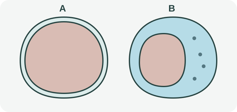
Original schematic, not anatomy to scale or a medical image. A and B compare location only, not severity.
- A — Narrow pleural space
A small gap separates the illustrated lung and chest boundary.
- B — Air outside the lung
Blue depicts air in that space; the coloured lung occupies less of the cross section.
The air is in the pleural space, outside the lung
Pneumothorax means air has entered the space between the lung and chest wall, allowing part or all of the lung to collapse. It is not simply extra air in the airways, nor a synonym for mucus blockage, pneumonia or every form of a “collapsed lung”.
The conceptual drawing shows location, not a scan: the coloured lung is inside the outer chest boundary. In B, the blue region is air outside the lung, and the lung is smaller. The dots do not measure air volume, pressure or severity.
Spontaneous, primary and secondary describe different features
“Spontaneous” distinguishes an event without an identified trauma or procedure as its trigger. “Primary” describes occurrence without a known underlying lung disease; “secondary” describes occurrence with an underlying lung disease. These labels do not classify a person’s symptoms.
Primary does not mean that every aspect of the lung has been proved normal. It is therefore misleading to interpret the word as “no possible connection with smoking”. Conversely, a smoking history does not by itself turn a pneumothorax into a secondary one.
Recurrence begins with people who already had an episode
A first-occurrence comparison asks who develops pneumothorax in an initially eligible population. A recurrence comparison starts after an episode. Gloucestershire Hospitals’ current leaflet says people who continue smoking after primary spontaneous pneumothorax are more likely to have another.
Illustrative comparison: a follow-up report lasting one year and another lasting five years give different opportunities for recurrence to be counted. A group receiving one form of care also cannot automatically stand for every other group. A recurrence percentage needs its population and follow-up period; it is not a personal forecast.
Bring the actual question to the clinical team
An NHS leaflet is written for its own patients and includes clinical follow-up instructions. Its discussion can explain terms, but individual follow-up, work, exercise, flights or diving need the responsible clinician’s advice. This page sets none of those schedules.
A useful question is: “Does this figure concern a first episode or another episode, and over what period?” Smoking-support services can explain available support separately. General guidance on smoking does not guarantee that pneumothorax will never recur.
What to keep in mind
Sources
The central claims on this page were checked against the sources below.
- NHLBI / NIH: Pleurisy, Pleural Effusion, and Pneumothorax
Sources checked: 2026-10-10
- NHLBI / NIH: Pleural Disorders: Causes and Risk Factors
Sources checked: 2026-10-10
- Gloucestershire Hospitals NHS Foundation Trust: Spontaneous primary pneumothorax
Sources checked: 2026-10-10
- Österreichisches Gesundheitsportal: Pneumothorax
Sources checked: 2026-10-10
Adult general education. No symptom diagnosis, imaging interpretation, procedure selection or individual activity and travel clearance.